Stem Cell Models of Embryonic Development and Human Organogenesis in a Dish
The mission of our laboratory is to unlock the mysteries of human early development for improvement of reproduction and regeneration biology.
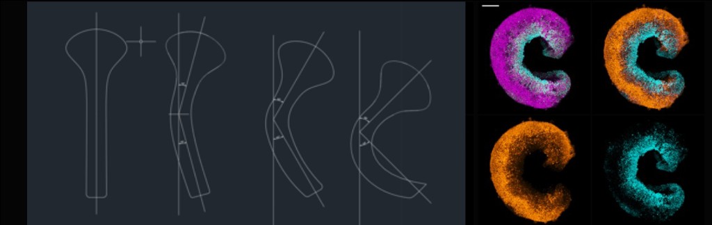
We focus on gastrulation and early organogenesis stages, a critical period during which tissue and organ rudiments are specified and patterned, however remains as a block box in human development.
We primarily work on three questions:
How we tackle the above questions
"Our real teacher has been and still is the embryo, who is, incidentally, the only teacher who is always right" — Embryologist and developmental neuroscientist Victor Hamburgers (1900-2001)
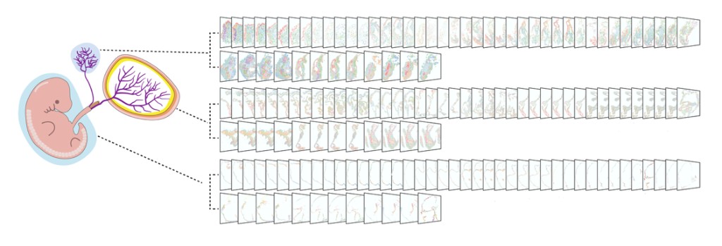
We learn how embryo develops from the real teacher, the embryo itself. We first collect spatial multi-omics data across embryonic stages to gain insights into how developmental programs unfold in vivo. This value dataset not only allows us to decode natural embryogenesis but also provides essential blueprint and benchmark for recapitulating aspects of embryos using stem cells in vitro.
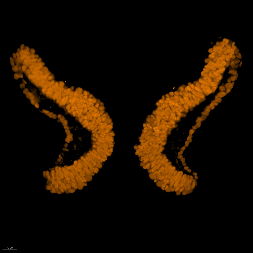
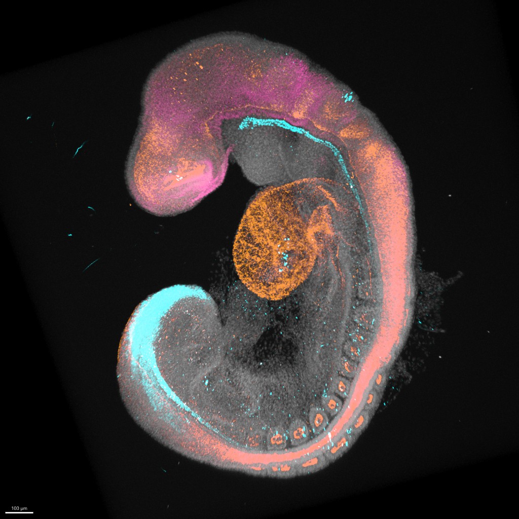
We apply high resolution and quantitative imaging to examine how the germ layers and organ rudiments are generated, and what can cause developmental abnormalities, in both animal models and stem cell-based embryo models (SCBEMs).
With the aid of in vivo knowledge at an unprecedented scale, including RNA, protein and metabolomics information, we harness the developmental potential and self-organization ability of pluripotent stem cells to mimic aspects of natural embryos, particularly those of humans. Those so-called stem-cell-based embryo models (SCBEMs) are powerful tools that permit the dissection of molecular and cellular mechanisms underlying body axis formation, germ layer specification and morphogenesis during the "black box" period of human development. This period is otherwise challenging to study due to limited access to human samples and ethical constrains.

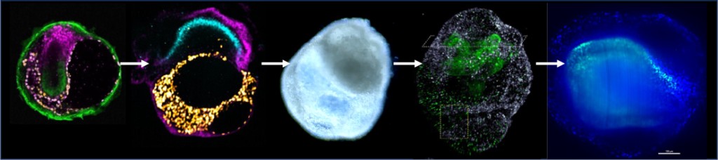
We also adopt other new technologies which enable experiments not feasible using traditional ways. For instance, we use bioengineering methods like micropatterning and 3D printing to manipulate tissue geometry and signaling gradients. Those allow us to investigate how those factors influence cell-cell communications and cell fate patterning.